
Medical Disclaimer: This article is AI-assisted and published strictly for general educational purposes in accordance with the National Medical Commission (NMC) guidelines. It does not establish a doctor-patient relationship and does not replace professional medical advice, diagnosis, or treatment. Please consult a qualified healthcare specialist for evaluation specific to your condition.
Quick Summary for Parents & Caregivers
A choledochal cyst in a child is a rare congenital anomaly marked by abnormal enlargement or cystic dilation of the bile ducts. Common symptoms include jaundice (yellow skin/eyes), persistent abdominal pain, pale/clay-colored stools, and a palpable lump in the upper right abdomen. The primary cause is an Anomalous Pancreaticobiliary Junction (APBJ). Surgical removal of the cyst followed by biliary reconstruction (Roux-en-Y Hepaticojejunostomy) is the mandatory definitive treatment to prevent recurrent infections, liver damage, and future malignancy. Seek expert pediatric care from Dr. Sujit Chowdhary for specialized Choledochal Cyst Treatment in Delhi.
Discovering that your child or infant has been diagnosed with a choledochal cyst can be an overwhelming experience for any family. Parents often hear terms like biliary dilatation, pancreaticobiliary maljunction, or Roux-en-Y reconstruction and feel anxious about what these medical labels mean for their child's health and future quality of life.
Understanding the anatomy of the liver and bile ducts, recognizing subtle warning signs early, and seeking guidance from an experienced pediatric surgeon are the most vital steps you can take. Fortunately, with modern advances in pediatric hepatobiliary surgery and minimally invasive laparoscopic techniques, choledochal cysts can be completely cured through surgical intervention, allowing children to grow up healthy, active, and fully vibrant.
In this comprehensive guide, Senior Pediatric Urologist and Surgeon Dr. Sujit Chowdhary explains everything parents need to know about pediatric choledochal cysts—from underlying congenital causes and clinical symptoms to advanced diagnostic imaging and gold-standard surgical options for Choledochal Cyst Treatment in Delhi.
Seeking Expert Consultation for a Choledochal Cyst?
Consult with internationally trained pediatric surgeon Dr. Sujit Chowdhary for precise diagnosis and advanced laparoscopic surgical care.
Book an Appointment TodayWhat is a Choledochal Cyst in Children?
To understand what a choledochal cyst is, it helps to understand how the body produces and transports bile. The liver produces bile, a digestive fluid essential for breaking down fats and eliminating waste products from the bloodstream. Bile travels from the liver through a network of small tubes called bile ducts (the biliary tree), gets stored temporarily in the gallbladder, and eventually flows into the duodenum (the first part of the small intestine).
A choledochal cyst is a congenital structural defect where parts of the extrahepatic or intrahepatic bile ducts undergo abnormal, pouch-like expansion or cystic dilation. Instead of remaining a smooth, narrow pipe, the bile duct balloons out. This structural malformation causes bile to stagnate (biliary stasis), leading to severe inflammation, bile duct stone formation, recurrent bacterial infections, and progressive liver tissue injury if not treated promptly.
Key Fact for Parents
Choledochal cysts are congenital anomalies present at birth, though symptoms may manifest during infancy, early childhood, or occasionally remain silent until adolescence. Female children are affected approximately 3 to 4 times more frequently than male children.
Types of Choledochal Cysts (Todani Classification)
Pediatric surgeons classify choledochal cysts into five distinct categories based on their anatomical location and pattern of dilation, using the internationally accepted Todani Classification system:
- Type I (Solitary Extrahepatic Dilation): The most common form (accounting for 80% to 90% of all cases), involving cystic or fusiform enlargement of the common bile duct outside the liver.
- Type II (Extrahepatic Diverticulum): A rare pouch-like sac or diverticulum protruding outward from the wall of the common bile duct.
- Type III (Choledochocele): Dilation situated within the wall of the duodenum at the lower end of the bile duct near the Ampulla of Vater.
- Type IV (Multiple Dilation): Dilation affecting both intrahepatic (inside the liver) and extrahepatic (outside the liver) bile ducts.
- Type V (Caroli's Disease): Rare cystic expansion confined strictly to the intrahepatic bile ducts within the liver tissue.
What Causes a Choledochal Cyst in a Child?
While the exact genetic or developmental trigger is still being researched, the widely accepted medical explanation for pediatric choledochal cysts centers on an anatomical defect known as an Anomalous Pancreaticobiliary Junction (APBJ) or pancreaticobiliary maljunction.
1. Anomalous Pancreaticobiliary Junction (APBJ)
In normal human anatomy, the common bile duct and the main pancreatic duct join together just as they enter the duodenal wall, sharing a short common channel controlled by a sphincter mechanism (the Sphincter of Oddi). In children born with an APBJ, these two ducts unite prematurely outside the intestinal wall, creating an abnormally long common channel.
Because the pressure inside the pancreatic duct is higher than inside the bile duct, pancreatic digestive juices continuously flow backward (reflux) into the bile duct. These active pancreatic enzymes digest and erode the delicate elastic fibers of the bile duct wall. Over time, this chemical irritation weakens the duct wall, causing it to dilate and swell into a choledochal cyst.
2. Congenital Structural Weakness & Obstruction
In some cases, localized stenosis (narrowing) or dysmotility at the distal end of the common bile duct increases internal fluid pressure, contributing to ballooning of the proximal bile duct above the obstruction.
Symptoms of Choledochal Cyst in Children
The symptoms of a choledochal cyst can vary significantly depending on the child's age, cyst size, degree of bile obstruction, and whether secondary bacterial infections or pancreatic inflammation have developed.
| Age Group | Primary Clinical Symptoms | Common Diagnostic Indicators |
|---|---|---|
| Infants & Newborns | Neonatal jaundice, pale/clay-colored stools (acholic stools), dark urine, abdominal distension, failure to thrive. | Persistent conjugated hyperbilirubinemia, cystic mass visible on infant ultrasound. |
| Toddlers & Children | Recurrent right upper quadrant abdominal pain, intermittent jaundice, fever with chills (cholangitis), nausea, vomiting. | Elevated LFTs, acute pancreatitis flares, palpable abdominal mass. |
| Adolescents | Vague abdominal discomfort, indigestion, recurrent gallstone/bile stone symptoms, unexplained jaundice. | Biliary sludge, elevated amylase/lipase, ductal wall thickening. |
The Classic Diagnostic Triad
In classical medical textbooks, choledochal cyst is described by a triad of three key symptoms:
- Jaundice: Yellowing of the whites of the eyes (sclera) and skin due to impaired bile drainage.
- Abdominal Pain: Episodic or constant pain located in the right upper quadrant of the stomach.
- Palpable Mass: A smooth, firm lump or swelling felt by the doctor in the upper right abdomen.
Note: In modern clinical practice, only 20% to 30% of pediatric patients exhibit all three symptoms simultaneously. Many children present with only one or two isolated signs, or the cyst is discovered incidentally during routine abdominal sonography for unrelated issues.
Warning Signs Parents Should Never Ignore
- Pale or Clay-Colored Stools: When bile cannot enter the intestine, stool loses its natural brown color and turns chalky white, grey, or pale yellow.
- Dark Tea-Colored Urine: Excess bilirubin filtered by the kidneys stains the urine dark yellow or brownish-tea color.
- Recurrent Unexplained Fever with Chills: Indicates bacterial cholangitis (infection of stagnant bile inside the cyst), which is a serious medical emergency requiring urgent IV antibiotics.
- Unexplained Abdominal Pain & Vomiting: May signify acute pancreatitis resulting from pancreatic juice reflux.
How is a Choledochal Cyst Diagnosed?
Accurate diagnostic evaluation is essential to confirm the presence of a choledochal cyst, delineate its precise anatomical structure, evaluate the pancreatic junction, and plan the surgical approach.
1. Antenatal / Prenatal Ultrasound
With widespread use of routine maternal ultrasound scans during pregnancy, many choledochal cysts are now detected in utero during the second or third trimester as a fluid-filled cystic mass under the fetal liver.
2. High-Resolution Abdominal Ultrasonography (USG)
Abdominal ultrasound is the primary non-invasive screening tool for pediatric liver and biliary tract conditions. It clearly visualizes cystic dilatation of the common bile duct, assesses gallbladder wall thickness, and evaluates intrahepatic bile duct involvement.
3. Magnetic Resonance Cholangiopancreatography (MRCP)
MRCP is the gold-standard imaging modality for choledochal cyst evaluation in children. It provides high-resolution 3D images of the entire biliary tree, hepatic ducts, and pancreatic duct junction without exposing the child to ionizing radiation or invasive dye injections.
4. Laboratory Blood Tests (Liver Function Tests)
- Serum Bilirubin: Measures total and direct (conjugated) bilirubin levels to assess bile duct blockage.
- Liver Enzymes: Elevated Alkaline Phosphatase (ALP), Gamma-Glutamyl Transferase (GGT), AST, and ALT indicate liver inflammation and biliary stasis.
- Serum Amylase & Lipase: Evaluated to check for co-existing pancreatitis.
Complications of Untreated Choledochal Cyst
Leaving a choledochal cyst untreated or delaying surgical intervention carries grave health risks for a child. Because stagnant bile is prone to bacterial colonization and chronic irritation, untreated cysts can lead to severe long-term complications:
- Recurrent Acute Cholangitis: Severe bacterial infection of the biliary tree causing high fever, sepsis, and liver abscess formation.
- Acute & Chronic Pancreatitis: Inflammation of the pancreas due to pancreatic duct reflux or stone obstruction.
- Biliary Cirrhosis & Portal Hypertension: Persistent bile backpressure causes progressive liver fibrosis, leading to irreversible liver failure over time.
- Cyst Rupture: Spontaneous perforation of an inflamed cyst wall leading to life-threatening peritonitis (abdominal lining infection).
- Malignancy (Cholangiocarcinoma): Long-term chronic inflammation in the cyst wall dramatically increases the risk of bile duct cancer later in adult life (up to 20-30% risk if left unexcised). This is why complete surgical removal of the cyst is mandatory.
Treatment Options for Choledochal Cyst in Children
There is no non-surgical or medical cure for a choledochal cyst. While medications like antibiotics or bile acid liquefiers can temporarily control active infections or jaundice, surgical excision is the only definitive treatment.
Expert Surgical Care for Your Child
Dr. Sujit Chowdhary specializes in advanced laparoscopic choledochal cyst excision and Roux-en-Y reconstruction with exceptional surgical outcomes.
Explore Choledochal Cyst Treatment DetailsThe Gold-Standard Surgical Procedure
The definitive, globally recognized surgical procedure for Type I and Type IV choledochal cysts consists of two essential steps performed during a single operation:
- Complete Excision of the Cyst & Gallbladder (Cholecystectomy): The surgeon meticulously dissects and removes the entire dilated cyst sac down to its junction with the pancreatic duct, along with the gallbladder. Complete removal of all abnormal cyst tissue is critical to eliminate future cancer risk.
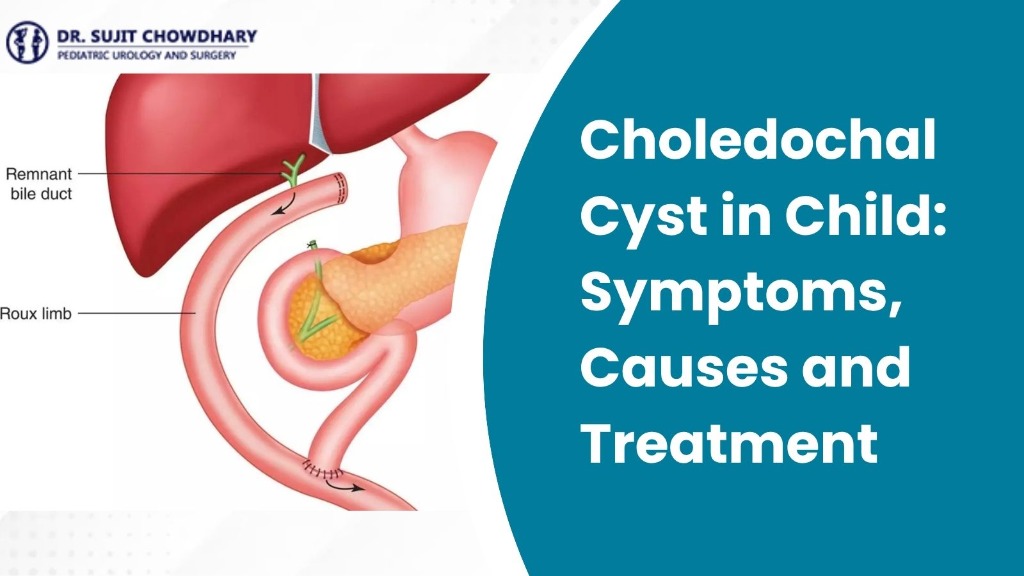
- Biliary Tree Reconstruction (Roux-en-Y Hepaticojejunostomy): To restore continuous bile flow from the liver into the digestive tract, a loop of the small intestine (jejunum) is brought up and cleanly attached directly to the healthy hepatic bile duct emerging from the liver. This reconstructed path allows bile to flow smoothly into the bowel without stagnation or pancreatic reflux.
Open vs. Laparoscopic (Keyhole) Choledochal Cyst Surgery
Traditionally, choledochal cyst excision was performed via an open abdominal incision. Today, in specialized pediatric centers offering expert Choledochal Cyst Treatment in Delhi under senior surgeons like Dr. Sujit Chowdhary, laparoscopic (minimally invasive keyhole) surgery is widely performed.
| Surgical Feature | Laparoscopic (Keyhole) Surgery | Traditional Open Surgery |
|---|---|---|
| Incision Size | 3–4 tiny keyhole punctures (3-5 mm) | Single large upper abdominal incision |
| Post-Operative Pain | Minimal pain, reduced pain medication requirement | Moderate to severe incisional discomfort |
| Hospital Stay | 3 to 5 days | 7 to 10 days |
| Cosmetic Outcome | Virtually invisible microscopic scars | Prominent permanent abdominal scar |
| Recovery Speed | Rapid return to normal feeding and play | Gradual recovery over several weeks |
Recovery and Long-Term Post-Surgical Care
After undergoing successful choledochal cyst excision and Roux-en-Y reconstruction, children recover remarkably fast. Within a few days, oral feeding is resumed, jaundice rapidly clears, and liver function normalizes.
Post-Operative Management Tips for Parents
- Dietary Transition: Start with light liquids, progressing smoothly to a normal balanced childhood diet rich in proteins and vitamins.
- Activity Restrictions: Encourage quiet indoor play for 2 to 3 weeks following surgery, avoiding heavy contact sports until full healing is confirmed.
- Routine Follow-Up Visits: Periodic liver function tests (LFTs) and abdominal ultrasounds are scheduled at 3 months, 6 months, and annually to ensure smooth bile passage and healthy liver growth.
With timely surgical repair, the long-term prognosis for children with a choledochal cyst is outstanding. Over 95% of children lead completely normal, active lives with no dietary limitations or growth delays.
Frequently Asked Questions (FAQs)
Q1. What is the main cause of a choledochal cyst in a child?
The primary cause is a congenital structural anomaly called an Anomalous Pancreaticobiliary Junction (APBJ). This abnormal connection allows high-pressure pancreatic digestive juices to flow backward into the main bile duct, eroding its walls and causing it to dilate into a cyst over time.
Q2. Can a choledochal cyst dissolve or heal on its own without surgery?
No. A choledochal cyst is an irreversible structural malformation of the bile duct walls. It cannot dissolve, shrink, or cure itself through medications or diet. Complete surgical excision is mandatory to prevent dangerous infections, liver cirrhosis, and long-term cancer risks.
Q3. At what age should a child undergo choledochal cyst surgery?
Surgery is typically recommended as soon as the diagnosis is confirmed and the child is medically stable. In infants diagnosed antenatally or shortly after birth, surgery is usually performed between 1 to 6 months of age to prevent early liver inflammation and cholangitis.
Q4. What is Roux-en-Y hepaticojejunostomy and why is it necessary?
Roux-en-Y hepaticojejunostomy is the surgical procedure used to reconstruct the biliary pathway after the diseased cyst and gallbladder are removed. A section of the small intestine (jejunum) is reshaped into a dedicated channel and connected directly to the liver's bile duct, allowing bile to drain safely into the digestive tract.
Q5. What are the warning signs of cholangitis in a child with a choledochal cyst?
Cholangitis is a bacterial infection of stagnant bile within the cyst. Key warning signs include sudden high fever with shaking chills, worsening jaundice (yellow eyes/skin), severe right upper abdominal pain, vomiting, and dark tea-colored urine. Cholangitis requires emergency hospital admission and IV antibiotic treatment.
Q6. Is choledochal cyst surgery safe for infants and young children?
Yes. When performed by an experienced pediatric hepatobiliary surgeon using modern pediatric anesthesia and laparoscopic keyhole techniques, choledochal cyst excision is extremely safe, highly effective, and associated with high long-term success rates.
Q7. Will my child have to follow a special diet for life after surgery?
No! Once the Roux-en-Y reconstruction heals and bile flow is established, children can digest fat and process normal foods just like any other child. No lifelong dietary restrictions are necessary.
Q8. Why choose Dr. Sujit Chowdhary for Choledochal Cyst Treatment in Delhi?
Dr. Sujit Chowdhary is a internationally renowned Senior Consultant in Pediatric Urology & Surgery with over three decades of clinical expertise. He specializes in complex pediatric reconstructive surgery and laparoscopic Choledochal Cyst Treatment in Delhi, offering world-class care and exceptional patient outcomes.
Conclusion
A choledochal cyst diagnosis in a child requires prompt attention, accurate diagnostic imaging, and expert surgical planning. By understanding the symptoms, congenital causes, and mandatory role of surgical excision, parents can make informed, confident decisions for their child's healthcare journey.
With early intervention via complete cyst excision and Roux-en-Y hepaticojejunostomy reconstruction, children recover completely, enjoy normal digestions, and look forward to a healthy, bright future.
Consult Senior Pediatric Surgeon Dr. Sujit Chowdhary
If your child has been diagnosed with a choledochal cyst or exhibits unexplained jaundice and abdominal pain, get a comprehensive expert evaluation today.
Book Your Appointment Now